
Tracking Bugs and Trees, 2022.03.05
My excitement has been growing in regards to galls and cankers lately. Galls are abnormal growths or swelling induced by the interaction of an aggravating life form with the plant. Those life forms could be an insect, a mite, a fungi, bacterium, nematode, or even a virus. The plant releases growth hormones and chemicals to quickly form thick tissue growth around the animal, fungi, or bacteria which is interacting with the plant. Often you don’t even have to find the agent which causes the gall to know who it is. Instead you can examine the shape, location and host plant to determine who caused the gall to form. It’s another form of tracking, but this time you are finding the sign left behind by tiny things, instead of a Coyote (Canis latrans) or a bird.
Sort of similar to galls are cankers. Cankers are defined as necrotic lesions on a stem, branch, or twig of a plant. They can kill the plants, but not always. I have been curious about cankers for a while since noticing some interesting ones around Southern Ontario on various trees and shrubs, but only recently have I been trying to research them. When we went out tracking at Mono Cliffs last weekend I was watching as we went for any signs of galls and cankers.
The first gall we came across I was familiar with. A few years ago I attended a workshop on Goldenrod (Solidago and Euthamia spp.) galls with Emily Damstra, who is an amazing scientific illustrator. She elaborated on all sorts of different galls she had found on various Goldenrod species, all of which she had illustrated as well. One of the galls she had talked about was the Scarred Goldenrod Gall, which is caused by a moth, Epiblema scudderiana. I had confused this gall with a very similarly shaped gall caused by a different moth, Goldenrod Elliptical Gall Moth, otherwise known as Gnorimoschema gallaesolidaginis. The former isn’t as narrow, nor as regular in form. E. scudderiana’s galls are a bit lumpier and seemingly larger, at least according to the photographs I have. In the field between to the Bruce Trail and the forested area before the cliffs, there were plenty of American Crow (Corvus brachyrhynchos) tracks around the tall stalks of the Goldenrods. When the group got closer folks were noticing that the Crow tracks led right up to the broken stalks of the Goldenrods, where the galls had been split open and the contents (which would likely be a larva) were missing. It seems like the Crows are eating the moth larvae! How coool!! I have seen the work of Black-capped Chickadees (Poecile atricapilus) and Downy Woodpeckers (Picoides pubescens) pecking out the larva of Eurosta solidaginus, the Goldenrod Ball Gall Fly, from the round orb-like galls on the stalks of Goldenrods before, but I had not seen anyone else mowing down on the larvae of these moths. It was really exciting. I ended up carrying around the broken stalk with the busted gall for a while, and I took way too many photos just to make sure I got all the angles.

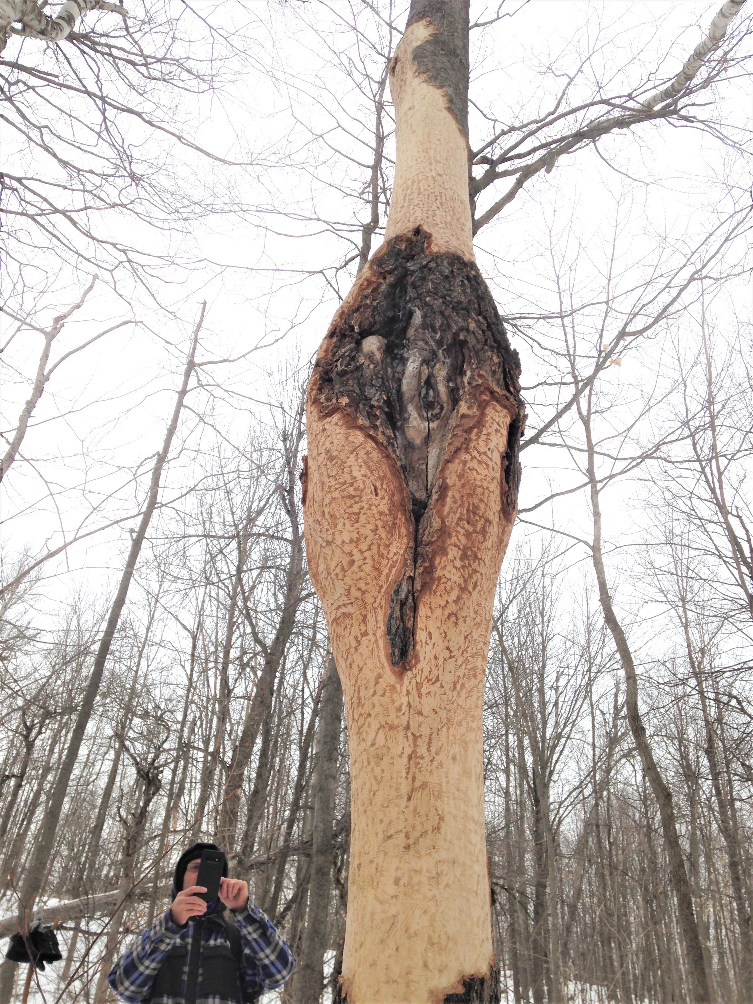
We continued on towards the direction of the forest. I knew from previous visits we would get the chance to see lots of big Sugar Maples (Acer saccharum) and some American Beech (Fagus grandifolia) and I was hoping to see some signs on them. As we moved through the forest edge, composed of some Apples (Malus domestica) and Trembling Aspen (Populus tremuloides), Alexis pointed out a couple of old wounds on the Aspens which could have been from White-tailed Deer (Odocoileus virginianus) antler rubs. I got to wondering if the wounds were from Eutypella or Nectria canker on the Aspens doing similar damage?
My research seems to be showing me that the Eutypella and Nectria canker doesn’t really show up on Aspens. It seems like this is just how the Aspen heals from the antler rub. It would make sense that the two forms of damage would look similarly because we aren’t just seeing the signs of the damage. We are actually seeing the signs of the trees sealing over the wound. In the image we can see two living lobes (callus, soft tissue from the cambium layer) on either side of exposed sapwood. These two lobes are slowly growing over top of the wound, and eventually they will meet and they will create a seam. In the following years the tree will continue to grow, out and up, and soon will cover up the seam with new growth and the old wound will be invisible from the outside. This sealing over is similar whether the damage is done by fungi, fire, or feisty buck. With this in mind, I think it would be a fun exercise to find a new rub somewhere near my home and continue to check on it year after year to document how the tree seals the wound.
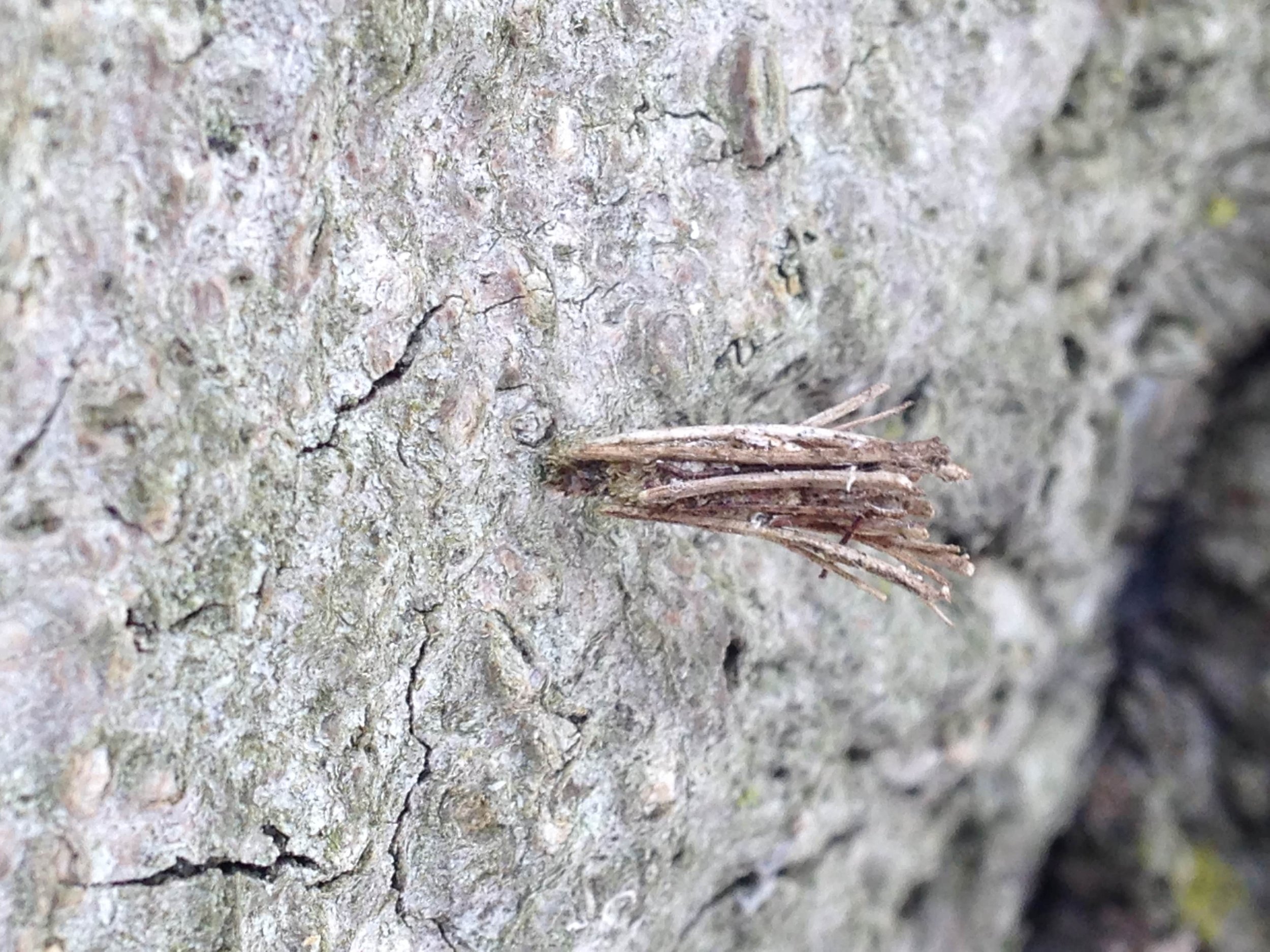
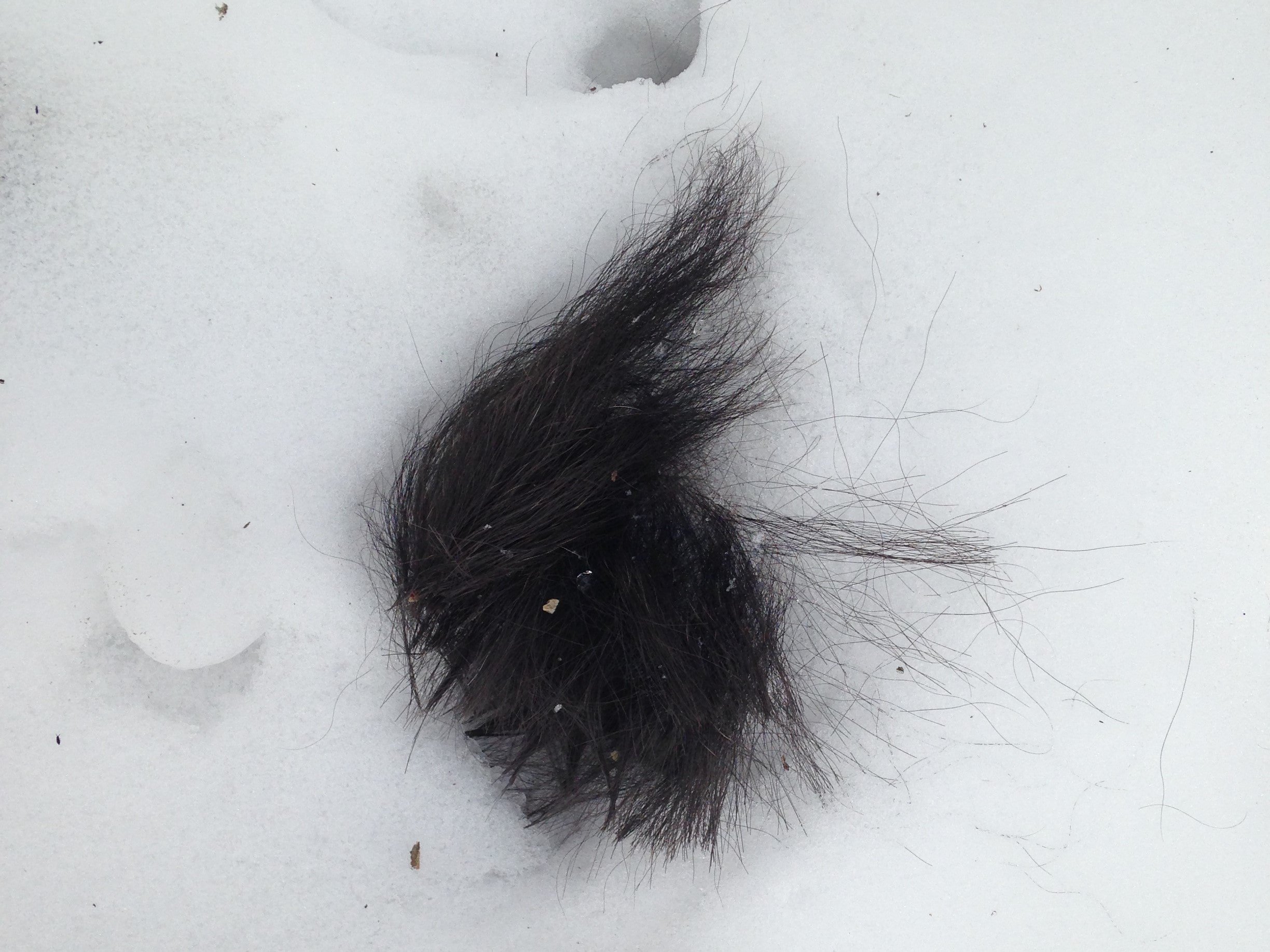
We checked out a few more tracks as we tucked into the deeper forest closer to the cliffs. I took a ton of photos, but none really showed too well in the very overcast day. The shadows we faint and the tracks hard to see in the shots I got. We did get to look at a couple of the cases of some Common Bagworm Moths (Psyche casta) left attached to a dead Beech tree which was a first for me. I had read about the Bagworm Moths but never had I seen one. There were also some tiny mystery insect eggs, 40 or so of them, strung along a fine thread wrapped around two branches. I still haven’t identified who they belong to but I do intend to. They were somewhat similar to Sawfly cocoons, though much smaller and so many together along that fine but strong strand. After that it looked like a Fisher (Pekania pennanti) was sniffing around at a black morph Gray Squirrel (Sciurus carolinensis) tail. We couldn’t decide whether the Squirrel had be eaten by the Fisher, or if the Fisher just happened upon the tail just sitting on top of the snow, but there was no blood, not a lot of hair, a couple of holes in the snow leading to Squirrel sized tunnels, but all those things together do not indicate a kill.
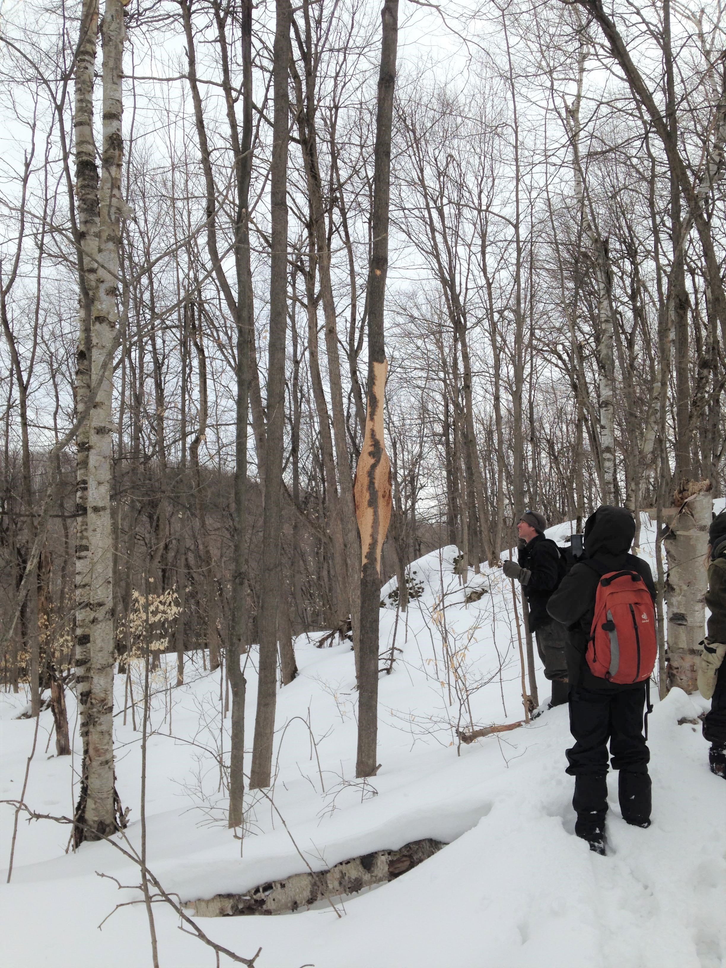
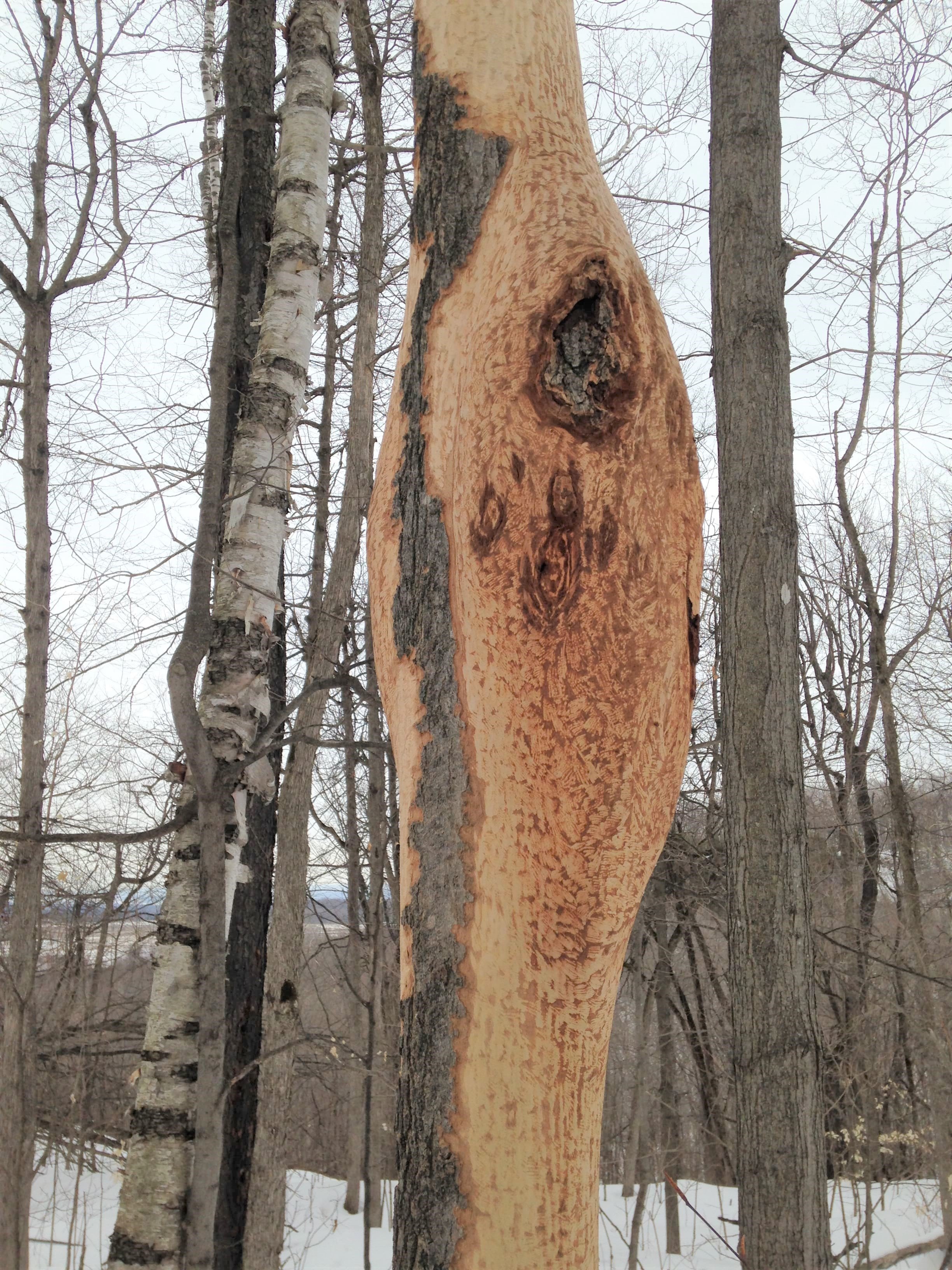

After the Fisher and the Squirrel sign, we noticed another large tree in the distance, seemingly glowing like a lighthouse in the forest. It was a Sugar Maple which had huge sections of bark gnawed off and eaten by a Porcupine (Erethizon dorsatum). This was cool in and of itself, but what really struck me was that the main area of the bark that the Porky had chewed off was on the backside of a flared “Cobra Canker” which is caused by Neonectria ditissima, or in one of my older books, Nectria galligena.
The way the Neonectria ditissima “Cobra Canker” happens is that the fungi gets in a damaged area of the tree and kills off some of the bark in the damaged area. The inner bark or “phloem” dries up and cracks, the bark sloughs off, and falls away from the tree. The tree host then creates a protective rim of callus around the canker (the “necrotic lesion” if we want to be metal about it). This is an effort to stop the spread and eventually allow the callus layer to seal up the damaged area. BUT, when the trees go dormant in Winter and they can’t fight back, the N. ditissima fungus kills the new callus layer. AND THEN, the tree wakes up in the Spring to this new damage all “what the hell?”-like, isolates the harm, and creates new callus lobes to seal over the area once again. BUT, when the trees go dormant in Winter… Slowly, year after year, the canker grows and new lobes are created. This eventually starts to look like the spread neck-flaps of a threatened King Cobras (Ophiophagus hannah). This canker doesn’t just affect the Sugar Maples. Other hosts include Large-toothed Aspen (Populus grandidentata), Trembling Aspen, Basswood (Tilia americana), Beech, Paper Birch (Betula papyrifera), Yellow Birch (Betula alleghaniensis), Red Maple (Acer rubrum), and some Willows (Salix spp.).
The way the Porcupine had gone at this Maple really had me full of questions. I got to wondering if the Maple was creating any sort of defence chemicals to deal with the canker, which might have the byproduct of being valuable to the Porcupine in some way? Does the Sugar Maple make more sugars in these areas? Does the new growth of callus tissue require extra minerals and proteins which make the bark extra beneficial? So far I can’t find any information on this, but it’ll be in my question book for the future.
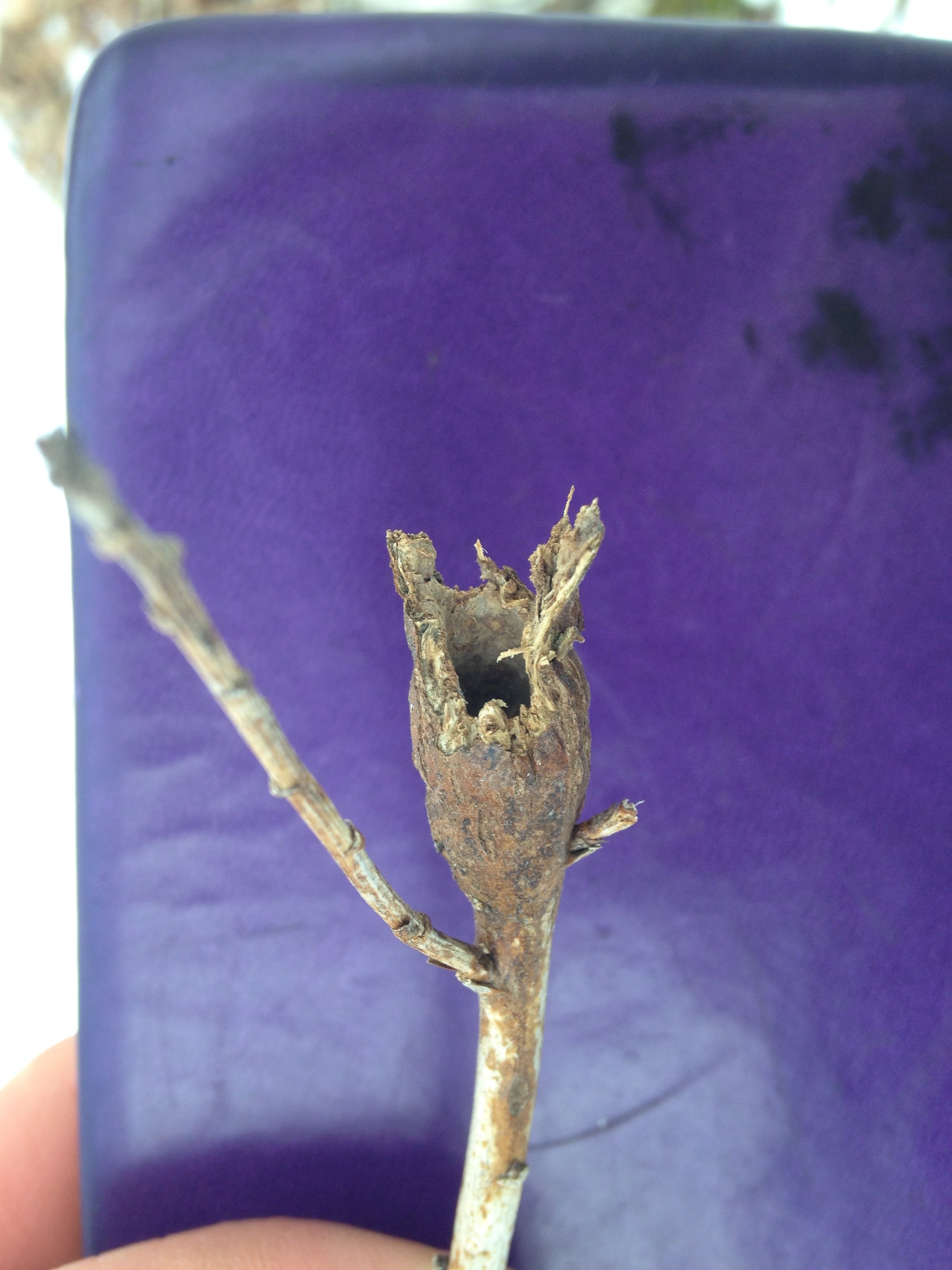
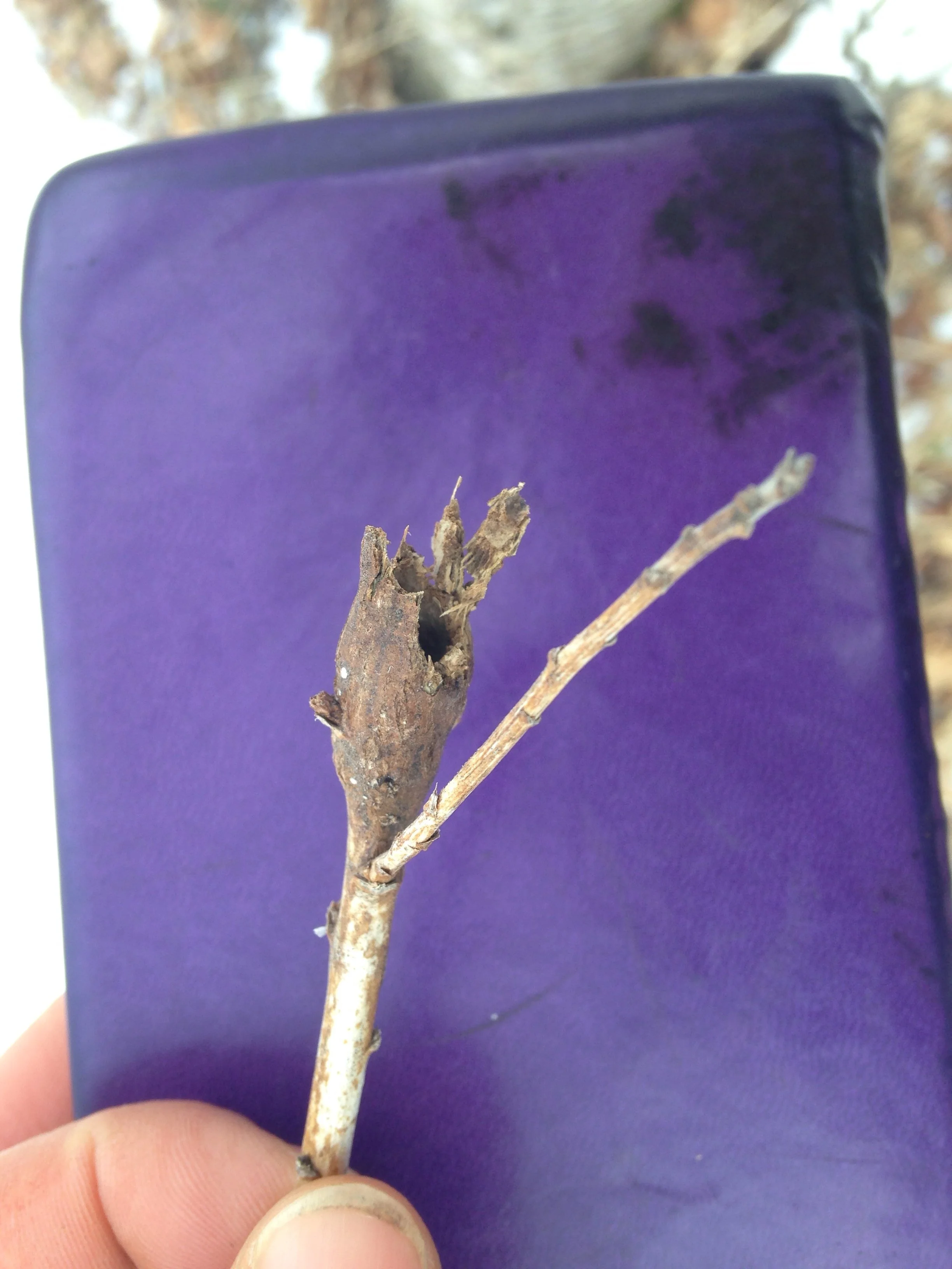
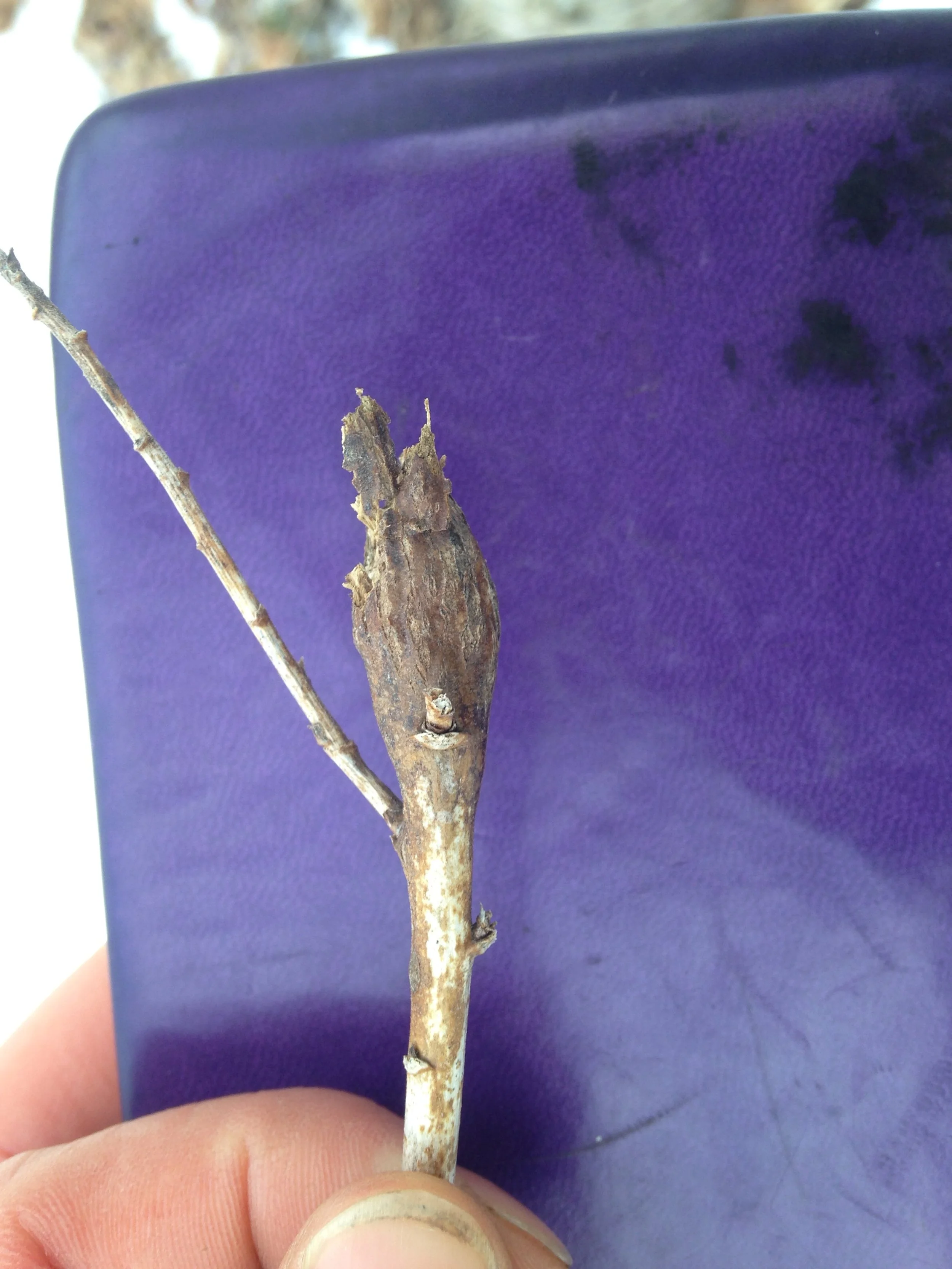
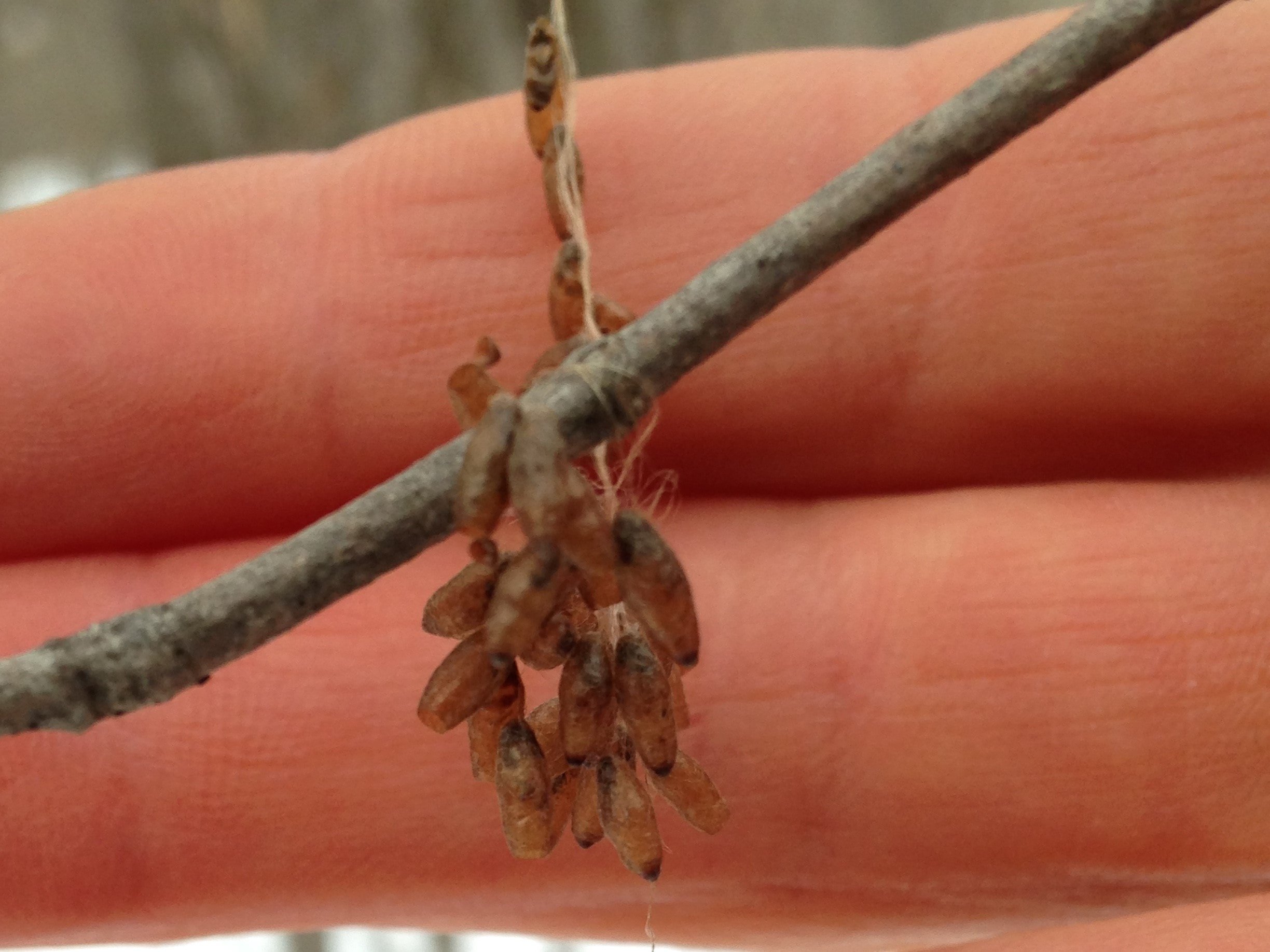
Further down the hill we began looking for a place to have lunch. While trailing behind the group I came across a few more signs of N. ditissima on Maples, but also saw something I have been investigating for the past few weeks. When I saw the bright red branches of the Red-Osier Dogwood (Cornus sericea) I stopped and looked, hoping that I would find the then-unknown woody gall growing mid-way up the branches. And to my luck I did find it. I ended up excitedly taking a couple of photos which turned out terribly so I’ve also included photos from another instance of when I came across this same gall.
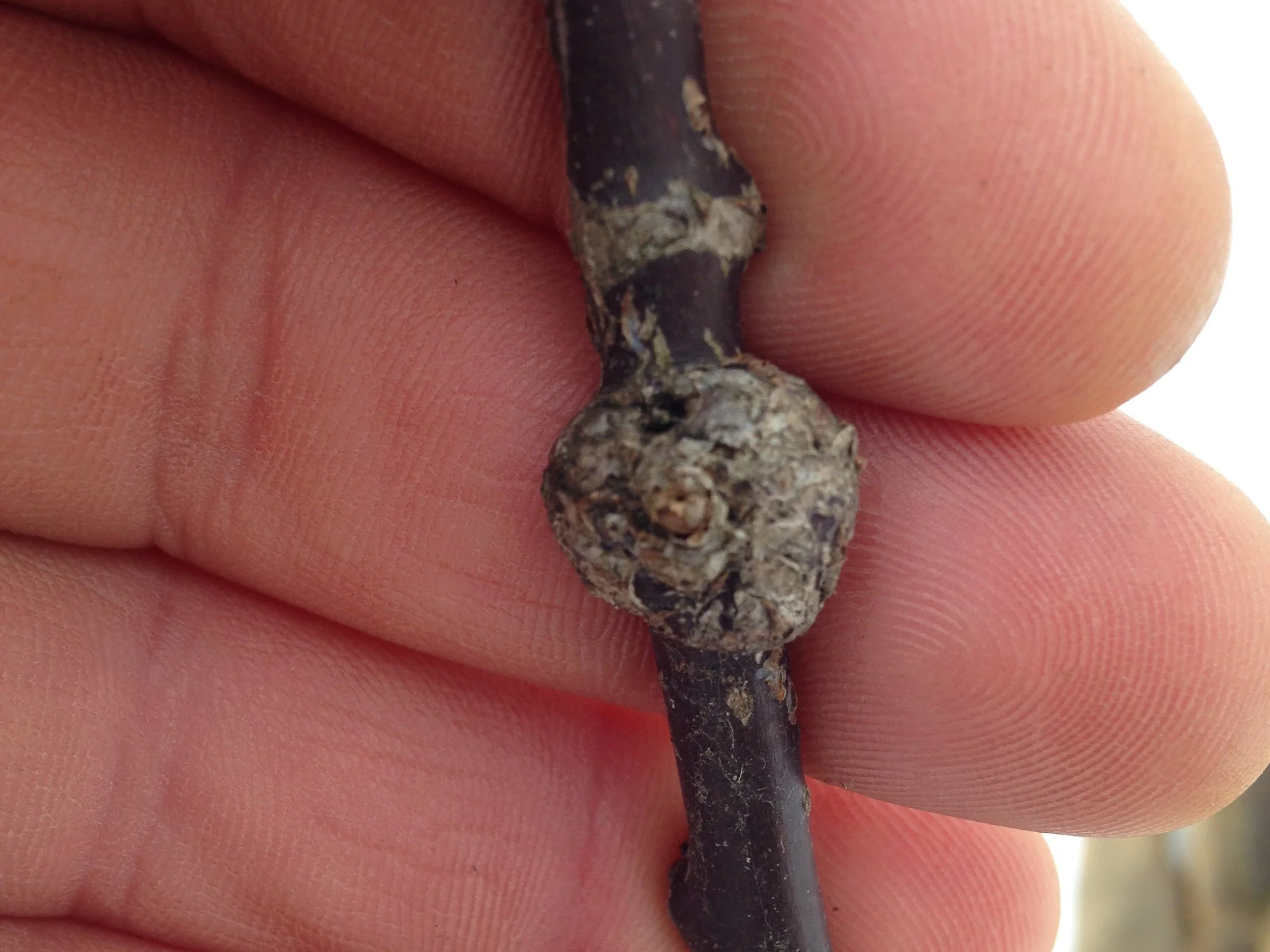
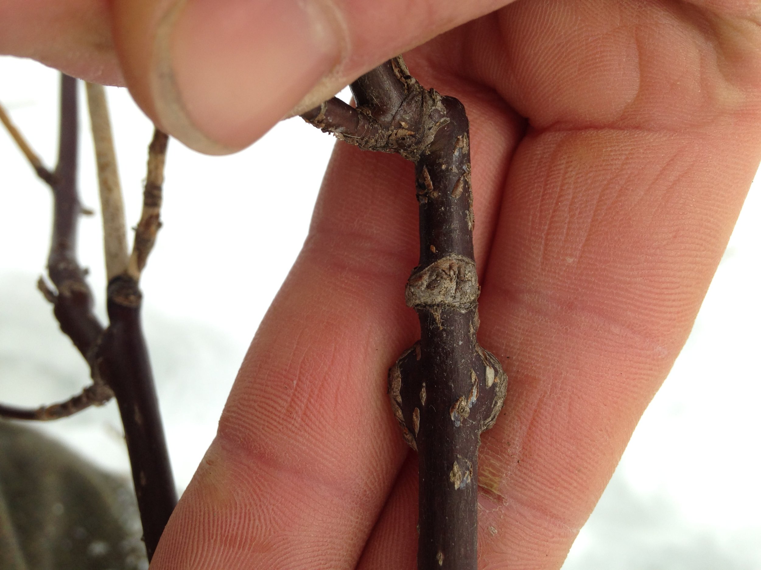
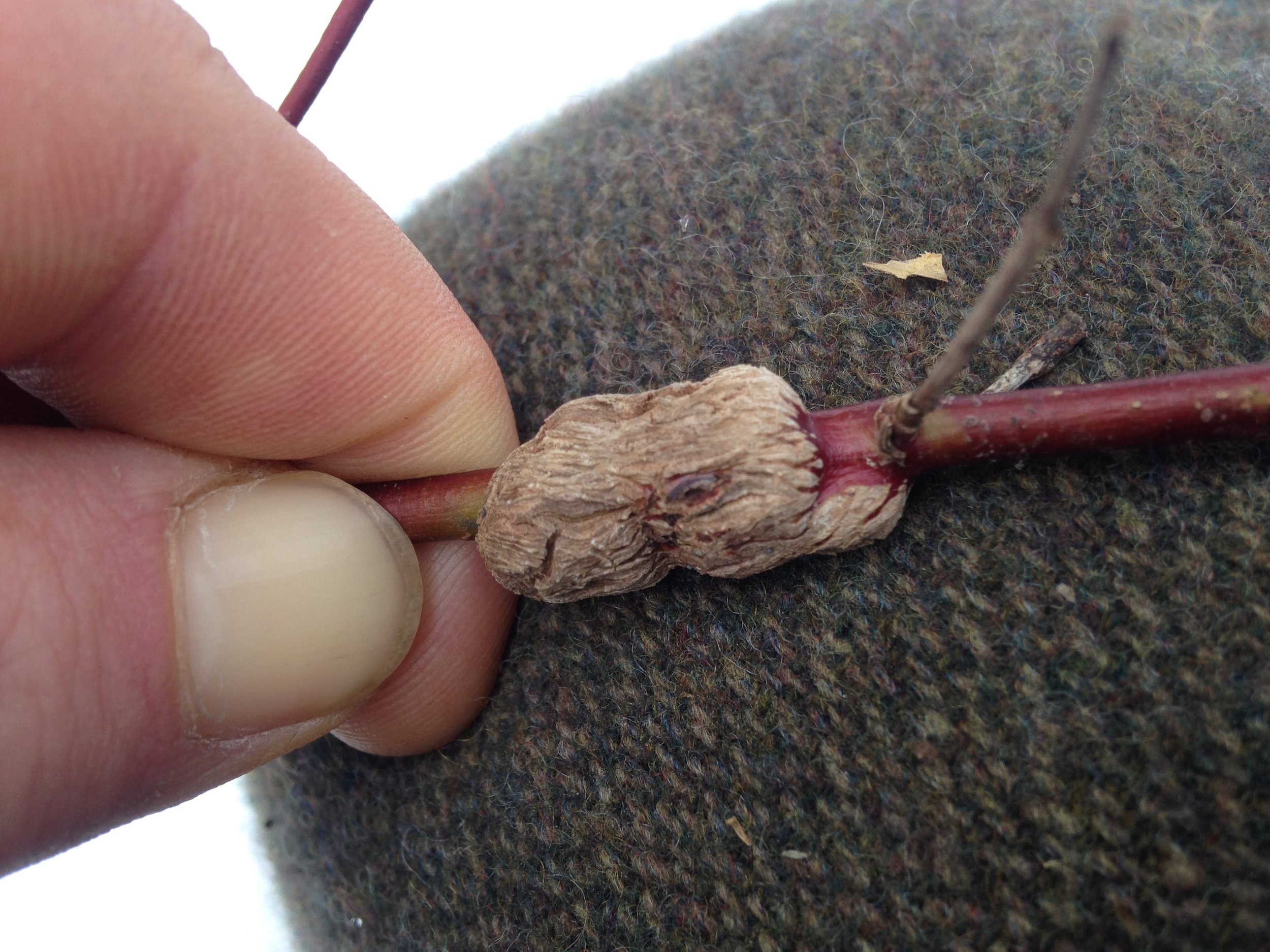
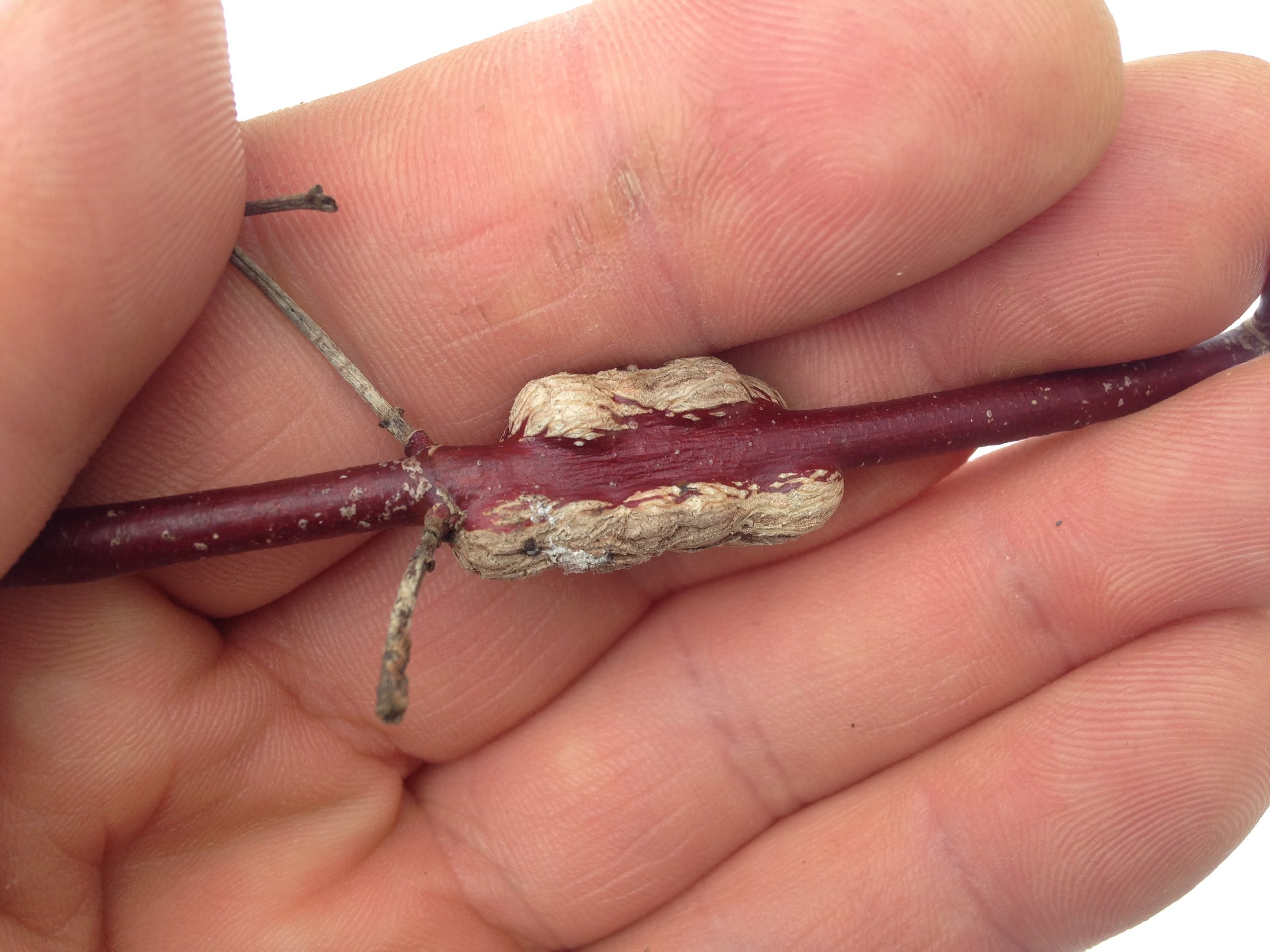
I had been trying to figure out who was making these galls for the past week, checking out all my books, looking at photos online, asking on message boards, all to no avail, until shortly after taking this photo I spoke with Alastair who mentioned a website about galls which he had just learned about, gallformers.org. I took it to heart and when I got home after that day of tracking and I looked up what I thought the gall might have been, the “Dogwood Club-gall” produced by a midge (a kind of fly) called Resseliella clavula. Just as I had suspected from my previous research, the Club-gall diagnosis appeared to be wrong. As I looked at other photos online of the Club-galls themselves it felt like I was forcing that i.d. as the shapes of the galls were pretty different. The Club-gall usually occurs at the tips of the branches, and this one was in the middle. The Club-galls are narrower at the base and then widen towards the top while this one was broad and lumpy throughout. There are often many Club-galls on a plant, but I was only finding one of these galls per plant. It just wasn’t right. I moped for half a second until I realized I could search by plant hosts. I looked up Cornus sericea and there was a single entry listed for Neolasioptera cornicola. I clicked the link and squealed! There was a range map, and an artists depiction of the gall I had found. I was overjoyed. The thrill of the hunt, the joy of the find.
Neolasioptera cornicola is a gall midge (also known as a gall gnat) which was described in the 1907 publication “New Species of Gall Producing Cecidomyiidae” by William Beutenmuller. The adult form of the insect described is pretty much a tiny black fly with a couple of pale bands around sections of the abdomen. The pale yellow larvae are described as kind of bumpy with a cone shaped head and reddish hind end. I was actually more interested in the description of the gall, which William describes as
Woody swellings on the twigs, branches or trunks on the dogwood (Cornus stolonifera*), measuring from about 6 to nearly 150 mm. in length. When situated on the twig, the gall is considerably smaller than those on the larger branches and trunk. It sometimes gnarls the trunk from about one to six inches or more in length.
(note that he uses the older name for the Red-Osier Dogwood, C. stolonifera, which is now known as C. sericea.)
I think I am going to have to keep looking for more of these Neolasioptera cornicola galls as I move through areas with C. sericea in hopes of finding some larger examples of the gall. Beutenmuller also wrote that the galls are common and can be found throughout all seasons of the year, making them something to be on the look out for. Lastly, he mentions that the larvae overwinter and pupate in the galls, and the adults emerge in May and June so this might be a great time to revisit all of the spots where I have seen the galls in hopes to watch one of the midges emerge. How cool would that be!?
While there was much more to the day in regards to track and sign of larger animals, like the drag marks from the remiges of a strutting Ruffed Grouse (Bonasa umbellus), taking the day to think about the interactions between tiny, even microscopic, life forms and the plant hosts they are in relationship with was a pretty damned special. These lifeforms inform larger communities and have ripples beyond their physical size. The Epiblema scudderiana moth which is food for the American Crow, or how the Sugar Maples and Paper Birches can become susceptible to blowdown in wind storms because the Neonectria ditissima canker weakens the trunk; these miniscule components of the broad forest ecology are both vital and nearly invisible in the woodlands where they live. To dig a little deeper and to understand these connections is really a joy and really inspiring. What other life is going on around us that we don’t know about and can barely see? How is the infinitely small making up and interacting with the immeasurably large? Who should I be asking? Where should I be looking? Life abounds, we just need to practice and hone skills to perceive it.
To learn more :
Emily Damstra’s website
Gallformers.org
Forest Pathology.org’s page on Target Cankers
Field Guide to Tree Diseases of Ontario - pdf
“New Species of Gall Producing Cecidomyiidae” by William Beutenmuller - pdf